- Double chambered right ventricle
- Hypertrophied muscle bundle (denoted by the star) can be seen in the right ventricular outflow tract causing obstruction
- Double chambered right ventricle
- Hypertrophied muscle bundle (denoted by the star) in right ventricular outflow tract
- Double chambered right ventricle
- Hypertrophied muscle bundle in right ventricular outflow tract (RVOT)
- Turbulent flow noted at the site of the muscle bundles in the RVOT
- Double chambered right ventricle
- Hypertrophied muscle bundle in right ventricular outflow tract denoted by the star
- Turbulent flow noted at the site of the muscle bundles in the RVOT
- Double chambered right ventricle
- Hypertrophied muscle bundle in right ventricular outflow tract denoted by the star
- Turbulent flow noted at the site of the muscle bundles in the RVOT
- Double chambered right ventricle
- Hypertrophied muscle bundle in right ventricular outflow tract denoted by the star
- Turbulent flow noted at the site of the muscle bundles in the RVOT
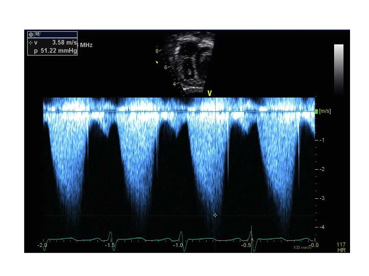
- Double chambered right ventricle
- Spectral Doppler across the right ventricular outflow tract in patient with double chambered right ventricle
- Moderate subpulmonary stenosis across a double chambered right ventricle
The subcostal short axis view is a excellent view to profile the right ventricular outflow tract and pulmonary valve. In cases of double chambered right ventricle, hypertrophied muscle bands can be visualized dividing the right ventricle into two chambers and causing right ventricular outflow tract obstruction. The severity of obstruction can be assessed from this view using color and spectral Doppler.
.png)
- Transducer on abdomen just below xyphoid process
- Notch at 6 o'clock
- Tilt the ultrasound probe leftward
- Double chambered right ventricle
- Pulmonary stenosis
- Supravalvar pulmonary stenosis