- Color compare view of main and branch pulmonary arteries
- Mild flow acceleration is noted starting in the proximal branch pulmonary arteries in a patient with mild bilateral PPS
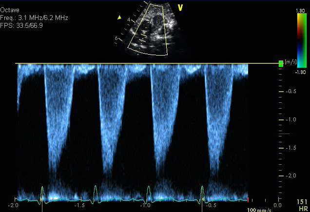
- Spectral pulse wave Doppler in the proximal right pulmonary artery
- Mild peripheral pulmonary stenosis (approximately 2 meters/sec)
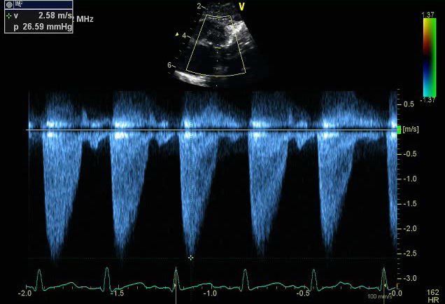
- Continuous wave spectral Doppler in the right pulmonary artery
- Mild peripheral pulmonary stenosis (~2.5 meters/sec)
The parasternal short axis is a good view to profile the size and flow pattern in the main and branch pulmonary arteries.
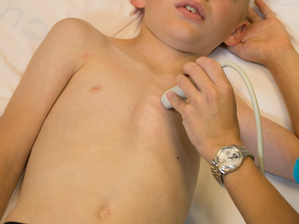
- Left sternal border
- 3rd or 4th intercostal space
- Notch pointing towards the left shoulder (1-2 o'clock)
- Transducer tilted superiorly and medially
- Pulmonary valve stenosis
- Supravalvar pulmonary stenosis
- Branch pulmonary artery stenosis