- Transitional AV Canal
- Large primum ASD
- Inlet VSD largely occluded by accessory AV valve tissue
- Moderate right atrial dilation
- Mildly dilated and hypertrophied right ventricle
- Transitional AV canal defect
- Moderate right AV valve regurgitation
- Mild to moderate left AV valve regurgitation
- Trivial amount of shunting through inlet VSD which appears largely occluded by accessory AV valve tissue (turbulent red color Doppler jet)
- Transitional AV Canal
- Moderate primum ASD with lett to right shunting
- Small inlet VSD largely occluded by accessory AV valve tissue with left to right shunting
- Moderate right AV valve regurgitation
- Mild to moderate left AV valve regurgitation
- Moderate right atrial dilation
Echocardiographic Assessment: Apical 4 Chamber
- Primum ASD (size and direction of shunting)
- Inlet VSD (size, assess for accessory AV valve tissue which often mostly or completely occludes VSD oriface)
- Right ventricle (dilation, hypertrophy, function)
- Left ventricle (dilation, hypertrophy, function)
- Atrioventricular valves (typicaly two orifaces)
- Right AV valve (stenosis or regurgitation)
- Left AV valve (stenosis or regurgitation)
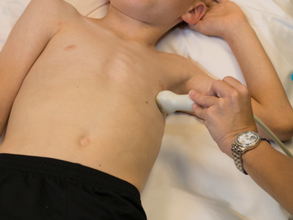
- Transducer placed on apical PMI (4th or 5th intercostal space)
- Midclavicular line at the apical PMI (point of maximal intensity)
- Notch at 3 o'clock