- PDA Constriction
- Moderately dilated and mildly hypertrophied right ventricle
- Moderately dilated right atrium
- Mild to moderately depressed right ventricular systolic function
- Small pericardial effusion
- PDA Constriction
- Small pericardial effusion
- Moderate tricuspid regurgitation
- Mild to moderately depressed right ventricular systolic function
- Moderately dilated right atrium
- Moderately dilated and mildly hypertrophied right ventricle
- PDA Constriction
- Moderately dilated and hypertrophied right ventricle
- Moderately dilated right atrium
- Moderately to severely depressed right ventricular systolic function
- Elevated RV pressures with bowing of the interventricular septum into the left ventricle
- PDA Constriction
- Moderately dilated and hypertrophied right ventricle
- Moderately dilated right atrium
- Moderate tricuspid regurgitation
- Moderately to severely depressed right ventricular systolic function
- Elevated RV pressures with compression of the left ventricle

- PDA Constriction
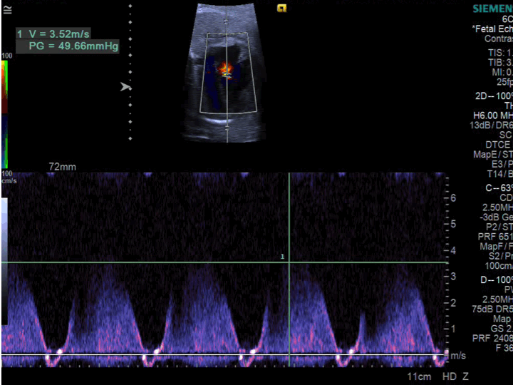
- Spectral Doppler of tricuspid regurgitation jet revealing elevated right ventricular systolic pressures
- PDA Constriction
- Flattened interventricular septal configuration with bowing of the interventricular septum (IVS) into the left ventricle consistent with elevated right ventricular systolic pressures
- Moderately to severely depressed right ventricular systolic function
- Hyperdynamic left ventricular systolic function
- PDA Constriction
- Restrictive and tortuous PDA with significant narrowing
- The star denotes the most narrow region
- PDA Constriction
- Restrictive and tortuous PDA with significant narrowing
- The star denotes the most narrow region
- Flow turbulence with continuous flow noted in constricted portion of PDA

- PDA Constriction
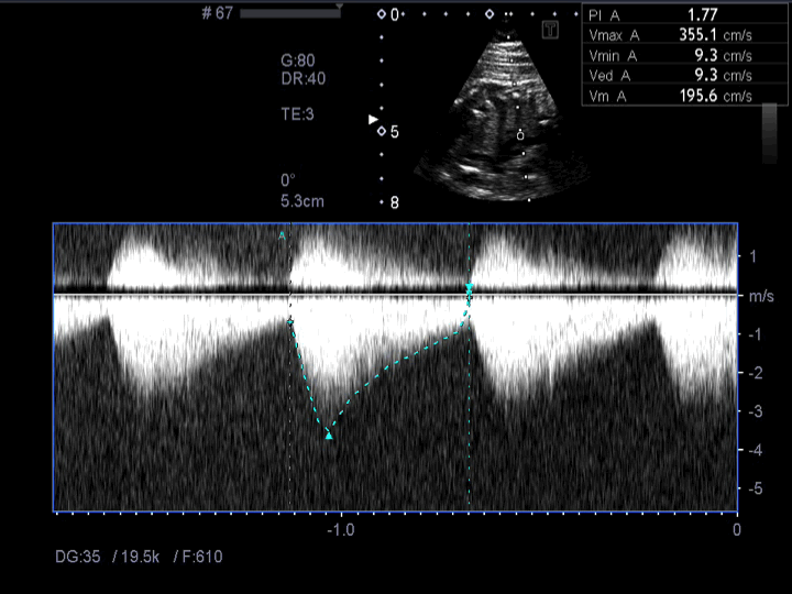
- Spectral Doppler in a PDA with ductal constriction
- Increased peak systolic (3.5 meters/sec) and diastolic velocities with a decreased pulsatility index (1.7)
- These findings meet criteria for ductal constriction
- PDA Constriction
- Sweep through aortic arch and PDA
- Restrictive and tortuous PDA with significant narrowing
- The star denotes the most narrow region
- Flow turbulence with continuous flow noted in constricted portion of PDA
- PDA Constriction
- Sweep through aortic and ductal arch
- Flow turbulence with continuous flow noted in constricted portion of PDA
- The star denotes the most narrow region
- Restrictive and tortuous PDA with significant narrowing with turbulent continuous flow in constricted PDA
- PDA Constriction
- Restrictive and tortuous PDA with significant narrowing
- PDA Constriction
- Flow turbulence with continuous flow noted in constricted tortuous portion of PDA
- Turbulent flow in constricted PDA
- PDA Constriction
- Sweep through aortic and ductal arch
- Restrictive and tortuous PDA with significant narrowing
- PDA Constriction
- Restrictive and tortuous PDA
- Color Doppler demonstrates tortuous PDA with flow turbulence

- PDA Constriction
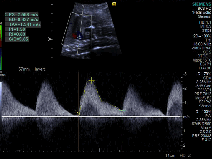
- Spectral Doppler in a PDA with ductal constriction
- Increased peak systolic (2.5 meters/sec) and diastolic velocities with a decreased pulsatility index (1.58)
- These findings meet criteria for ductal constriction