- Fetal Apical 4 Chamber View
- Normal fetal 4 chamber view
- Slight inferior displacement of the tricuspid valve compared to the mitral valve with chordal attachments from the tricuspid valve septal leaflet to the ventricular septum
- Smooth endocardial surface of the left ventricle
- Moderator band in the right ventricular apex
- Normal sized ventricles and AV valves
- Normal biventricular systolic function
- Echogenic focus in LV near mitral valve chordae/pappillary muscle (normal variant)
- Fetal Apical 4 Chamber View
- Normal fetal 4 chamber view
- Normal sized ventricles and AV valves
- Normal biventricular systolic function
- Fetal Apical 4 Chamber View
- Normal fetal 4 chamber view
- Normal sized ventricles and AV valves
- Color Doppler with normal laminar tricuspid and mitral valve inflow
- No tricuspid or mitral regurgitation
- Fetal Apical 4 Chamber View (outflow tract sweep)
- Normal fetal 4 chamber view
- Sweep towards outflow tracts
- Great arteries arise from appropriate ventricles (aorta arises from LV and pulmonary artery from RV at end of sweep)
- Normal crossing of great arteries
- Small pericardial effusion (likely physiologic)
- Fetal Apical 4 Chamber View (outflow tract sweep)
- Normal fetal 4 chamber view
- Sweep towards outflow tracts
- Great arteries arise from appropriate ventricles (aorta arises from LV and pulmonary artery from RV at end of sweep)
- Normal crossing of great arteries
- Fetal Apical 4 Chamber View (outflow tract sweep)
- Sweep towards outflow tracts with color Doppler
- Great arteries arise from appropriate ventricles (aorta arises from LV and pulmonary artery from RV at end of sweep)
- Normal crossing of great arteries
- Normal laminar flor across outflow tracts and semilunar valves
- No ventricular level shunting (fetal echo cannot rule out small VSDs)
- Fetal Apical 4 Chamber View (outflow tract sweep)
- Sweep towards outflow tracts
- Great arteries arise from appropriate ventricles (aorta arises from LV and pulmonary artery from RV at end of sweep)
- Normal crossing of great arteries
- No obvious ventricular level shunting (fetal echo cannot rule out small VSDs)
- Fetal Apical 4 Chamber View (pulmonary veins)
- Two right and one left pulmonary vein seen returning normally to the left atrium
- Fetal Apical 4 Chamber View (pulmonary veins)
- Zoomed up view of left atrium
- Two right and two left pulmonary veins seen returning normally to the left atrium
- Zoomed up view of left atrium
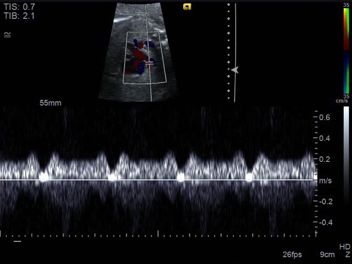
- Fetal Apical 4 Chamber View (pulmonary veins)
- Normal spectral Doppler pattern in a fetal pulmonary vein
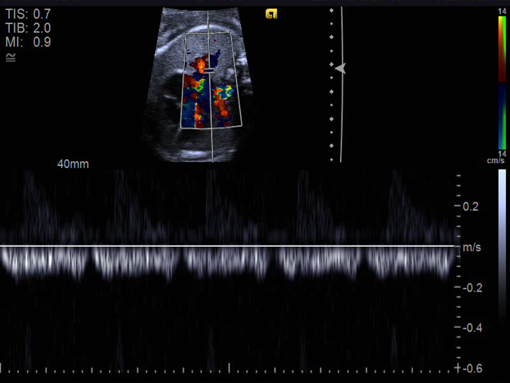
- Fetal Apical 4 Chamber View (pulmonary veins)
- Normal spectral Doppler pattern in a fetal pulmonary vein
- Fetal Apical 4 Chamber View (arch sidedness)
- Fetal apical 4 chamber view sweep towards outflow tracts
- The pulmonary artery and aorta arise normally from their respective ventricles
- At the end of the sweep the aorta is seen leftward of the trachea confirming a left-sided fetal aortic arch
These are normal fetal echo clips from an apical 4 chamber view.