- Partial AV Canal
- Moderate sized primum ASD
- Mild right atrial and right ventricular dilation
- Intact inlet ventricular septum with no evidence of an inlet VSD
- Partial AV Canal
- Moderate primum ASD with left to right shunting
- Mild mitral regurgitation via a cleft in the anterior mitral valve leaflet
- Mild tricuspid regurgitation
- Intact ventricular septum with no evidence of an inlet VSD
- Partial AV Canal
- Moderate primum ASD with left to right shunting
- Mild mitral regurgitation (via a cleft in the anterior mitral valve leaflet)
Echocardiographic Assessment: Apical 4 Chamber
- Primum ASD (size, direction of shunting)
- Right atrial dilation (secondary to left to right shunt across primum ASD)
- Left atrial dilation (secondary to regurgitation from mitral cleft)
- Right ventricular dilation (secondary to left to right shunt across primum ASD)
- Tricuspid valve (stenosis, regurgitation)
- Mitral valve (cleft, regurgitation, stenosis)
- Ventricular septum (assess for any evidence of inlet VSD)
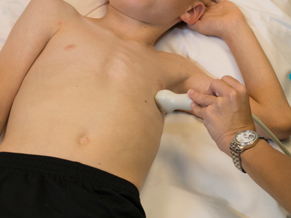
- Transducer placed on apical PMI (4th or 5th intercostal space)
- Midclavicular line at the apical PMI (point of maximal intensity)
- Notch at 3 o'clock