- Pulmonary Artery Sling
- Main pulmonary artery is seen which branches into the right pulmonary artery.
- There is no left pulmonary artery arising from the main pulmonary artery.
- Left pulmonary artery seen arising from the right pulmonary artery and branches leftward.
- Pulmonary Artery Sling
- Main pulmonary artery is seen which branches into the right pulmonary artery.
- There is no left pulmonary artery arising from the main pulmonary artery.
- Left pulmonary artery seen arising from the right pulmonary artery and branches leftward.
- Pulmonary Artery Sling
- Main pulmonary artery is seen which branches into the right pulmonary artery.
- There is no left pulmonary artery arising from the main pulmonary artery.
- Left pulmonary artery seen arising from the right pulmonary artery and travels posteriorly and leftward.
- Pulmonary Artery Sling
- Left pulmonary artery seen arising from the right pulmonary artery and travels posteriorly and leftward.

- Pulmonary Artery Sling
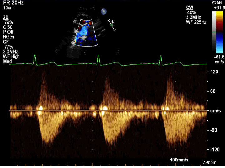
- Continuous wave Doppler in the left pulmonary artery
- The left pulmonary artery is unobstructed with low velocity pulsatile flow
Echocardiographic Assessment: High Parasternal Short Axis (Branch Pulmonary Arteries)
- By 2-D, measure the MPA, LPA, and RPA dimensions in systole.
- By 2D and color doppler, sweep to follow the LPA course as it goes posterior to the echo-bright trachea towards the left.
- Spectral doppler parallel to direction of flow to determine if there is any obstruction at the MPA, RPA, or LPA (perform both pulse wave and continuous wave doppler interrogation of MPA, proximal and distal RPA, proximal and distal LPA). Spectral doppler is especially important after LPA sling repair as these patients have a high incidence of LPA stenosis after repair.

- Modified parasternal short axis view
- Left sternal border
- 3rd or 4th intercostal space (slide upward 1-2 rib spaces and slightly more medial to obtain this modified view)
- Notch pointing towards 3 o'clock