- Pulmonary Artery Sling
- Main pulmonary artery is seen branching into the right pulmonary artery.
- There is no left pulmonary artery visualized from the main pulmonary artery
- Left pulmonary artery seen arising from the right pulmonary artery and going branching leftward.
- Pulmonary Artery Sling
- Main pulmonary artery is seen which branches into the right pulmonary artery.
- There is no left pulmonary artery by color seen arising from the main pulmonary artery.
- Left pulmonary artery arising from proximal right pulmonary artery and branches posteriorly and leftward.
- Pulmonary Artery Sling
- Main pulmonary artery is seen which branches into the right pulmonary artery.
- There is no left pulmonary artery by color arising from the main pulmonary artery.
- Left pulmonary artery arising from the right pulmonary artery and branches posteriorly and leftward.
- PDA with bidirectional shunting
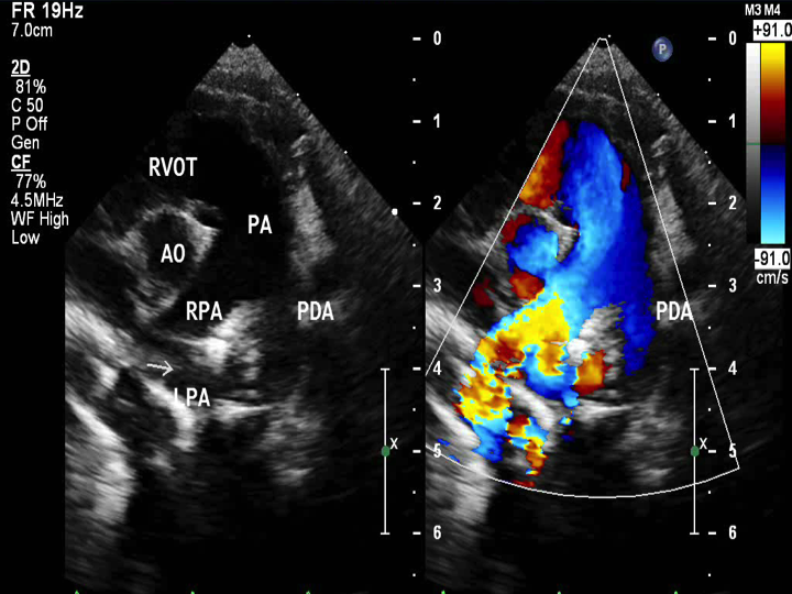
- Pulmonary Artery Sling
- Main pulmonary artery is seen which branches into the right pulmonary artery.
- There is no left pulmonary artery arising from the main pulmonary artery.
- Left pulmonary artery arising from the right pulmonary artery and branches posteriorly and leftward.
- What appears to be the left pulmonary artery is actually a PDA entering the main pulmonary artery
Echocardiographic Assessment: Parasternal short-axis pulmonary artery:
- Demonstrate the origin of left pulmonary artery (LPA) displaced to the right and arising from the posterior aspect of the right pulmonary artery (RPA)
- By 2-D, measure the main pulmonary artery (MPA), LPA, and RPA dimensions in systole
- By 2D and color doppler, sweep to follow the LPA course as it goes posterior to the echo-bright trachea and towards the left. Be careful not to mistake a patent ductus arteriosus (PDA) for the LPA as it may be along the same course of where the LPA would be expected (use color and spectral doppler to help you differentiate between these two structures).

- Left sternal border
- 3rd or 4th intercostal space
- Notch pointing towards the left shoulder (1-2 o'clock)
- Transducer tilted superiorly and medially