- Double Inlet Left Ventricle (left-handed ventricular topology, L-TGA)
- Apical view demonstrating left AV valve chordae seen beneath the pulmonary valve and crossing the VSD into hypoplastic subaortic outlet chamber
- The pulmonary valve appears thickened with tethered leaflets
- Double Inlet Left Ventricle
- Apical view demonstrating two AV valves aligned and connected to a single morphologic left ventricle with fine apical trabeculations
- Double Inlet Left Ventricle (right-handed ventricular topology, D-TGA)
- Apical view demonstrating parallel great arteries
- The aorta appears rightward and arises from the small outlet chamber
- Double Inlet Left Ventricle (right-handed ventricular topology, D-TGA)
- Apical color Doppler demonstrating parallel great arteries
- The aorta appears rightward and arises from the small outlet chamber
- Trivial aortic regurgitation
- Double Inlet Left Ventricle (right-handed ventricular topology, D-TGA)
- Apical sweep demonstrating two AV valves connected to the single left ventricle
- The pulmonary artery arises from the left ventricle (is seen as the bifurcating vessel)
- The aorta appears rightward and arises from the small outlet chamber (seen briefly at end of sweep)
- Double Inlet Left Ventricle (right-handed ventricular topology, D-TGA)
- Color compare view
- Left atrial enlargement
- Flow turbulence across left AV valve
- Small restrictive atrial communication with small amount of left to right shunting
- Findings overall suggestive of left atrial hypertension and a restrictive ASD
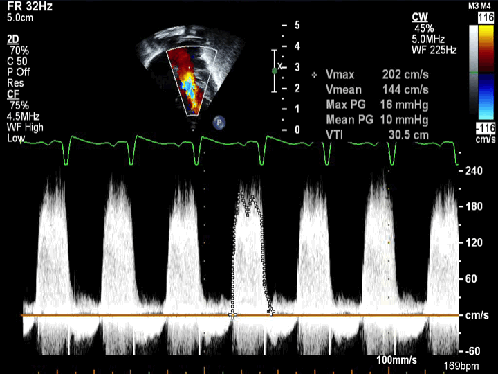
- Double Inlet Left Ventricle
- Continuous wave Doppler across the left AV valve with left AV valve stenosis with a mean gradient of 10 mmHg
- Patient with left atrial hypertension with left AV valve stenosis and a restrictive ASD
- Double Inlet Left Ventricle (right-handed ventricular topology, normally related great arteries, aka Holmes heart)
- Apical sweep demonstrating two AV valves aligned and connected to the single left ventricle
- Sweeping anteriorly demonstrates normally related great arteries
- The aorta arises posteriorly from the single left ventricle
- The pulmonary artery arises anteriorly (at end of sweep) from small right sided anterior outlet chamber
- Double Inlet Left Ventricle (right-handed ventricular topology, normally related great arteries, aka Holmes heart)
- Apical color Doppler sweep demonstrating two AV valves aligned and connected to the single left ventricle
- Sweeping anteriorly demonstrates normally related great arteries
- The aorta arises posteriorly from the single left ventricle
- The pulmonary artery arises anteriorly (at end of sweep) from small right sided anterior outlet chamber
- Double Inlet Left Ventricle
- Right and left AV valves both empty into a dilated left ventricle
- Double Inlet Left Ventricle
- Right and left AV valves both empty into a dilated and trabeculated left ventricle
Apical 4 chamber view.