- Double Inlet Left Ventricle
- Subcostal color compare of the atrial septum revealing a small restrictive PFO with left to right shunting
- Double Inlet Left Ventricle
- Subcostal color compare of the atrial septum revealing a small restrictive PFO with left to right shunting

- Double Inlet Left Ventricle
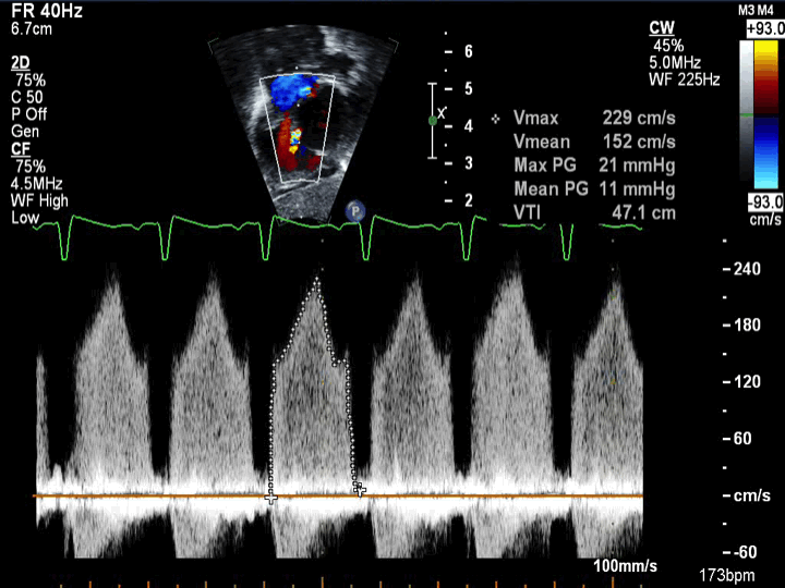
- Spectral Doppler across a PFO revealing restriction with an elevated mean gradient of 11 mmHg
-
DILV with normally related great arteries (D-looped/right-handed ventricular topology aka Holmes heart)
-
Subcostal color compare sweep demonstrating an anterior and rightward subpulmonary outlet chamber with normally related great arteries
-
Flow is seen into the hypoplastic RV chamber through a VSD
-
Pulmonary artery seen arising from hypoplastic right-sided RV chamber
-
Aorta seen arising from the left ventricle at the end of the sweep anteriorly (normally related great arteries)
-
- Double Inlet Left Ventricle (left-handed ventricular topology, L-TGA)
- Subcostal sweep demonstrating parallel great arteries with the aorta anterior and leftward (L-TGA)
- The anterior leftward aorta arises from a small anterior leftward subaortic hypoplastic RV cavity (seen at end of sweep)
- The pulmonary artery arises posteriorly from the LV
- There appear to be chordal attachments seen protruding into the subpulmonary region across the VSD
- Double Inlet Left Ventricle (left-handed ventricular topology, L-TGA)
- Subcostal color sweep demonstrating parallel great arteries with the aorta anterior and leftward (L-TGA)
- The anterior leftward aorta arises from a small anterior leftward subaortic hypoplastic RV cavity (seen at end of sweep)
- The pulmonary artery arises posteriorly from the LV
- There appears to be laminar flow across the VSD into the hypoplastic RV chamber and out the aorta (at end of sweep)
- There appear to be mild flow turbulence seen starting at the subpulmonary region near where chordal attachments are seen protruding beneath the pulmonary valve
- Double Inlet Left Ventricle (left-handed ventricular topology, L-TGA)
- Small anterior leftward subaortic hypoplastic RV cavity
- The pulmonary artery arises posteriorly from the LV
- AV valve chordae seen crossing the VSD
- Laminar flow across the VSD
- Mild flow turbulence if noted which begins in the subpulmonary region
Subcostal long axis view.