- Infracardiac total anomalous pulmonary venous return
- Descending vertical vein seen draining into the portal circulation
- Infracardiac total anomalous pulmonary venous return
- Vertical vein drains inferiorly into the portal vein/ductus venosus
- Turbulent flow noted at the insertion of the vertical vein into the portal vein/ductus venosus concerning for pulmonary venous obstruction
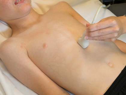
Echocardiographic Assessment: Subcostal IVC
- Vertical vein anatomy and course
- Assess by 2D, color and spectral Doppler for evidence of turbulence/obstruction
- most commonly as the vertical vein descends below the diaphram and enters the portal venous circulation
- Assess by 2D, color and spectral Doppler for evidence of turbulence/obstruction
- Inferior vena cava/portal vein/ductus venosus
- Size and flow by 2D, color and spectral Doppler
- Assess for connection to vertical vein
- Assess for obstruction as vertical vein connects to portal venous circulation

- Probe position in subxyphoid location
- Rotate probe so notch at 12 o'clock
- Tilt to patient's left