- Infracardiac total anomalous pulmonary venous return
- Four pulmonary veins seen draining to a confluence posterior to the left atrium.
- Infracardiac total anomalous pulmonary venous return
- At least three pulmonary veins are seen draining to a confluence posterior to the left atrium
- The venous confluence subsequently drains to a vertical vein which will ultimately descend inferiorly below the diaphraghm
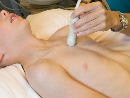
Echocardiographic Assessment: Suprasternal Notch
- Pulmonary venous confluence
- Assess individual pulmonary veins as they drain into confluence by 2D, color and spectral Doppler
- Vertical vein anatomy and course
- Assess origin and course by color and spectral Doppler for evidence of turbulence/obstruction
- Most common site of obstruction as vertical vein travels below the diaphragm and enters the portal venous circulation

- Probe at suprasternal notch
- Notch at 3 o'clock
- Transducer tilted posteriorly (tail up)
- May have to slide probe inferiorly along the left side of sternum with a posterior inferior tilt of the transducer (tail up) to fully profile pulmonary veins