- Aortopulmonary window
- Left atrial and left ventricular dilation in a patient with a large AP window
- Echocardiographic Assessment: Apical Four-Chamber View
- 2D: Evaluate the hemodynamic significance of shunting across the AP window by evaluating left atrial and left ventricular size.
- Apical 4-chamber and 2-chamber views should be performed to quantify extent of left atrial dilation.
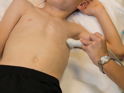
- Transducer placed on apical PMI (4th or 5th intercostal space)
- Midclavicular line at the apical PMI (point of maximal intensity)
- Notch at 3 o'clock