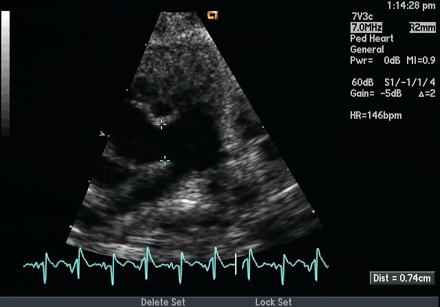
- Echocardiographic Assessment: Parasternal Short Axis Pulmonary Artery
- 2D: The main pulmonary artery (MPA) is seen giving off the branch pulmonary arteries and the rightwards aorta is seen en face above the level of the aortic valve. Evaluate for drop-out in the great arterial walls and for extension into the right pulmonary artery. The AP window communication can be appreciated between the MPA and the aorta. In this view, also document coronary artery origins as AP window can be seen with coronary anomalies.
- Color Doppler: It is important to perform color Doppler in this view to confirm that there is flow across this drop-out in the great arterial walls.
- Still: Measurements of the defect can be made in this view.

- Left sternal border
- 3rd or 4th intercostal space
- Notch pointing towards the left shoulder (1-2 o'clock)
- Transducer tilted superiorly and medially

