- Echocardiographic Assessment: Apical Four-Chamber View Pulmonary Valve
- 2D: Sweep anteriorly while in the apical 4-chamber view to bring in the pulmonary valve. In this view, the great arteries association, ventricular septum, pulmonary valve morphology, and right ventricular outflow tract can also be evaluated as AP window can be seen with associated CHD lesions (ie - VSD, TOF, TGA). Evaluate the location of the AP window defect, distance from both semilunar valves, extent/presence of the inferior and superior rims, and if involvement of the right pulmonary artery exists.
- Color Doppler: Perform color Doppler in this view to confirm that there is flow across the great arterial walls. Keep in mind that earlier in the neonatal period when pulmonary vascular resistance is high, shunting across this lesion may not yet be seen. Therefore, it is important to repeat echocardiography if concerns for AP window exist in the early neonatal period. Directionality of shunting can also be appreciated in this view, and will commonly be bidirectional.
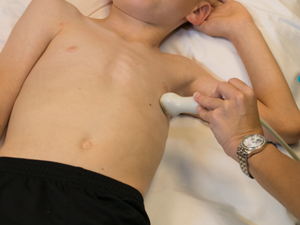
- Transducer at cardiac apex
- Notch at 3 o'clock
- Probe tilted very anteriorly (tail down)
- Sliding probe medially will optimize this view