- Cor Triatriatum Sinister
- Left atrial membrane visualized in mid portion of the left atrium
Echocardiographic Asssessment: Apical 2 Chamber
- Assess for presence of left atrial membrane and membrane morphology (location, size, single opening/fenestrated by 2D and color Doppler)
- Assess mean gradient across the membrane (pulse and continuous wave spectral Doppler)
- Left atrial size
- Mitral valve (stenosis, regurgitation by 2D, color and spectral Doppler)
- Pulmonary venous return into chamber above membrane (2D and spectral Doppler)
- Left atrial size
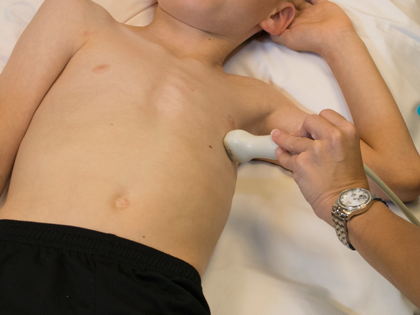
- Modified apical four chamber view
- From four chamber view rotate probe counter clockwise
- Indicator at 1 o'clock
- Transducer at mid clavicular line
- 4th or 5th intercostal space