- Cor Triatriatum Sinister
- A left atrial membrane is visualized in the mid portion of the left atrium
- Cor Triatriatum Sinister
- Left atrial membrane visualized in mid portion of left atrium
- Flow can be seen across the mid portion of the membrane
- The left lower pulmonary vein seen entering superior to the membrane (blue color Doppler jet)
- Cor Triatriatum Sinister
- Left atrial membrane visualized in the mid portion of the left atrium
- Cor Triatriaum Sinister
- Left atrial membrane visualized in mid portion of left atrium
- Turbulent color Doppler flow noted across a restrictive left atrial membrane
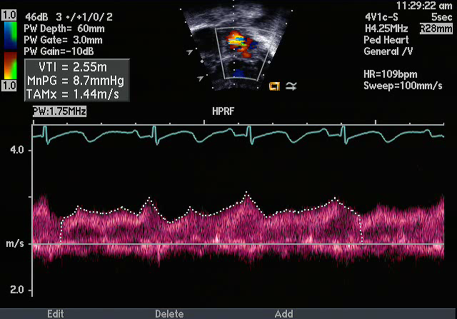
- Cor Triatriatum Sinister
- Spectral Doppler pattern across a restrictive left atrial membrane revealing stenosis across the membrane with a mean gradient of ~8.7 mmHg
- Cor Triatriatum Sinister
- Complex, thick and echogenic left atrial membrane dividing left atrium with two components to the membrane
- The starred cavity is the pulmonary venous chamber of the left atrium
- Severely dilated right ventricle with severe systolic dysfunction
- Frequent atrial ectopy with runs of atrial tachycardia
- Cor Triatriatum Sinister
- Complex, thick and echogenic left atrial membrane dividing left atrium
- Severely dilated right ventricle with severe systolic dysfunction
- Moderate tricuspid regurgitation
- Frequent atrial ectopy with runs of atrial tachycardia
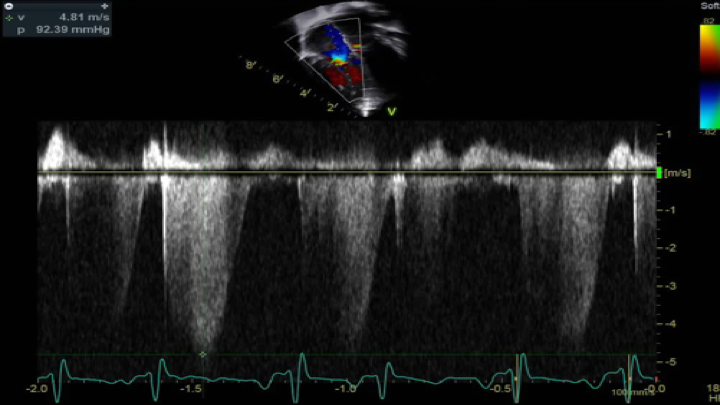
- Cor Triatriatum Sinister
- Tricuspid regurgitation spectral Doppler envelope
- The peak velocity is 4.8 meters/sec with a peak gradient of 92 mmHg + right atrial pressure which in this patient estimates right ventricular pressures at suprasystemic levels (secondary pulmonary hypertension in setting of pulmonary venous obstruction secondary to restrictive cor membrane)
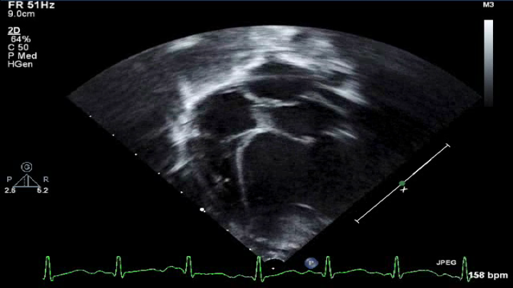
- Cor Triatriatum Sinister
- Apical 4 chamber still image of a Cor triatriatum membrane within the left atrium
- Cor Triatriatum Sinister
- Complex, thick and echogenic left atrial membrane dividing the left atrium
- Restrictive membrane with flow turbulence noted across at least two small fenestrations in the membrane
- Moderate tricuspid regurgitation
- Severely dilated right ventricle with severe systolic dysfunction
- Frequent atrial ectopy with runs of atrial tachycardia
- Cor Triatriatum Sinister
- Complex, thick and echogenic left atrial membrane dividing left atrium with two components
- Restrictive cor membrane with two to three small fenestrations with turbulent flow (represented by the arrows)
- At least two pulmonary veins are seen entering the pulmonary venous portion of the left atrium
- Severely dilated right ventricle with severe systolic dysfunction
- Frequent atrial ectopy with runs of atrial tachycardia
Echocardiographic Assessment: Apical 4 Chamber
- Assess for presence of left atrial membrane and membrane morphology (location, size, single opening/fenestrated by 2D and color Doppler)
- Assess mean gradient across the membrane (pulse and continuous wave spectral Doppler)
- Left atrial size
- Mitral valve (stenosis, regurgitation by 2D, color and spectral Doppler)
- Pulmonary venous return into chamber above membrane (2D and spectral Doppler)
- Assessment for pulmonary hypertension (tricuspid regurgitation by color and spectral Doppler with TR jet to assess RV systolic pressures)

- Transducer placed on apical PMI (4th or 5th intercostal space)
- Midclavicular line at the apical PMI (point of maximal intensity)
- Notch at 3 o'clock