- Tetralogy of Fallot
- Large conoventricular VSD (denoted by the star)
- The aorta overrides the large VSD and native ventricular septum
- Mild right ventricular hypertrophy
- Tetralogy of Fallot
- Large conoventricular VSD with left to right shunting
- The aorta overrides the large VSD and native ventricular septum
- Mild right ventricular hypertrophy

- Tetralogy of Fallot
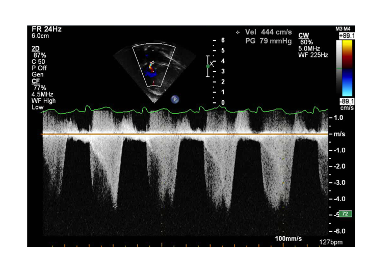
- Spectral Doppler across the right ventricular outflow tract (RVOT) and pulmonary valve from apical 5 chamber view (anteriorly)
- D= Dagger-like Doppler pattern consistent with dynamic obstruction across the RVOT secondary to infundibular narrowing
- PV= Second superimposed Doppler pattern consistent with fixed obstruction across the pulmonary valve.
- Spectral Doppler across the right ventricular outflow tract (RVOT) and pulmonary valve from apical 5 chamber view (anteriorly)

- Tetralogy of Fallot
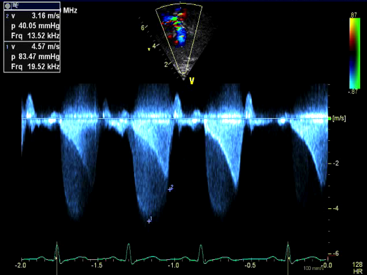
- Apical view tilted anteriorly into the RVOT and pulmonary valve revealed severe fixed and dynamic obstruction
- Two Doppler envelopes are noted
- Dagger-like signal consistent with dynamic (represented by D label) RVOT infundibular obstruction
- Well rounded Doppler signal consistent with fixed pulmonary valve stenosis (represented by PV label)