- Coarctation of the aorta
- Dilated right ventricle
- Moderate right and left ventricular systolic dysfunction
The apical 4 chamber view is a good view to assess for atrial and ventricular size and ventricular function in a patient with coarctation of the aorta.
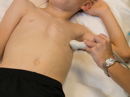
- Transducer placed on apical PMI (4th or 5th intercostal space)
- Midclavicular line at the apical PMI (point of maximal intensity)
- Notch at 3 o'clock
- Right or left atrial dilation
- Right or left ventricular dilation
- Right or left ventricular dysfunction
- Mitral valve hypoplasia or dysplasia