- Complete AV Canal
- Common AV valve equally committed emptying into both ventricles
- Moderate sized primum ASD
- Moderate sized inlet VSD
- Ventricles appear of normal size and are well balanced
- Mild RVH
- Mild right atrial dilation
- Complete AV Canal
- Common AV valve equally committed emptying into both ventricles
- Small primum ASD
- Large inlet VSD
- Mild right ventricular dilation and hypertrophy
- Complete AV Canal
- Zoomed view of complete balanced AV canal
- Common AV valve equally committed to both ventricles
- Small primum ASD
- Moderate inlet VSD
- Complete AV Canal
- Primum ASD with left to right shunting
- Mild right AV valve regurgitation
- Mild left AV valve regurgitation
- Moderate right atrial dilation
- Inlet VSD (shunting by color Doppler poorly profiled in this clip)
- Common AV valve appears balanced opening equally over both ventricles
- Balanced appearing ventricles without hypoplasia
- Complete AV Canal
- Zoomed color compare view of a complete balanced AV canal
- Small primum ASD with left to right shunting
- Moderate inlet VSD with bidirectional shunting
- Mild right AV valve regurgitation
- Mild left AV valve regurgitation
Echocardiographic Asssessment: Apical 4 Chamber
- Primum ASD (size and direction of shunting)
- Inlet (size and direction of shunting, peak velocity across VSD)
- Common AV valve (morphology, balanced versus unbalanced, stenosis, regurgitation)
- Ventricles (hypertrophy, dilation, function, balanced versus unbalanced)
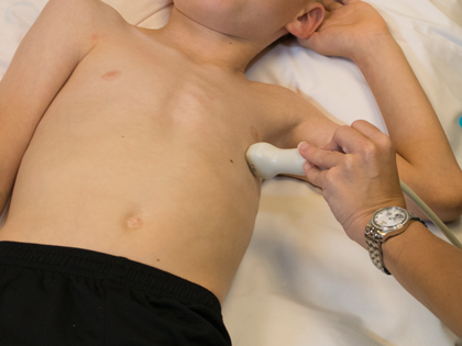
- Transducer placed on apical PMI (4th or 5th intercostal space)
- Midclavicular line at the apical PMI (point of maximal intensity)
- Notch at 3 o'clock