Complete Atrioventricular Canal (aka AV canal, common atrioventricular canal, atrioventricular septal defect, endocardial cushion defect)
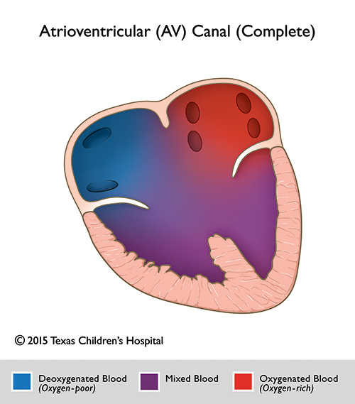
A complete atrioventricular canal (CAVC) describes a constellation of defects which comprise abnormalities in the structures that are derived from the endocardial cushions. In a complete AV canal, both the right and left atrium connect to the ventricles via a common (single) AV valve instead of two discrete (tricuspid and mitral) AV valves. There are deficiencies in both the atrial (primum ASD) and ventricular (inlet VSD) septum. CAVC results in a significant interatrial and interventricular systemic-to-pulmonary shunting, leading to right ventricular pressure and volume overload and pulmonary hypertension. CAVC accounts for approximately ~3% of cardiac malformations. Both sexes are equally affected and there is a significant association with Down syndrome/Trisomy 21. Patients often become symptomatic in infancy due to congestive heart failure and failure to thrive. Pharmacologic therapy (digoxin, diuretics, vasodilators) play a role as a bridge toward surgery which is usually performed between the 3-6 months of life.
Depending on the morphology of the superior leaflet of the common atrioventricular valve, 3 types of CAVC have been delineated (type A, B and C, according to Rastelli's classification). The anatomic subgroups (Rastelli's type A, B and C) can be classified on the basis of the chordal insertions and morphology of the superior bridging leaflet of the common atrioventricular valve.
- Rastelli Types
- Type A: The superior bridging leaflet demonstrates chordal attachments to the crest of the interventricular septum (~50-70%)
- Type B: The superior bridging leaflet attaches past the crest of the ventricular septum to an anomalous papillary muscle on the right side of the ventricular septum (~3%, rare)
- Type C: The superior bridging leaflet demonstrates no attachments to the ventricular septum and is referred to as a free floating leaflet (~30%).