- Echocardiographic Assessment: Parasternal Long Axis
- Profile the distal right ventricular outflow tract/subvalvar region (2D, color Doppler, spectral Doppler)
- Assess the pulmonary valve for dysplasia (leaflet morphology, excursion, hypoplasia, tethering)
- Assess main pulmonary artery (hypoplasia, post-stenotic dilation in case of valvar PS)
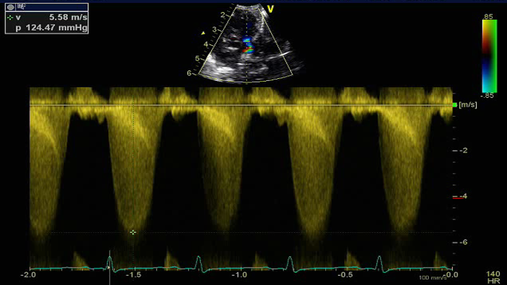
- Assess for pulmonary stenosis or regurgitation (color and spectral Doppler pulse wave and continuous wave Doppler) at the subvalvar, valvar and supravalvar level
.png)
- Transducer placed at the left sternal border
- 3rd or 4th intercostal space
- Notch pointed towards patient's right shoulder
- Angle transducer anteriorly towards patient's left shoulder (tilt tail of transducer down)