- Pulmonary Stenosis
- Severe pulmonary valve stenosis
- Dysplastic pulmonary valve with doming leaflets in systole
- The valve leaflets are severely restricted with a minimal amount of opening during systole
- Dilation of the main pulmonary artery
- Severe pulmonary valve stenosis
- Pulmonary Stenosis
- Severe pulmonary stenosis
- Trivial amount of prograde flow across the pulmonary valve (narrow turbulent blue Doppler jet at the central portion of the pulmonary valve in systole)
- Patent ductus arteriosus arteriosus (PDA) with left to right flow into the main pulmonary artery (turbulent red color Doppler jet)
- Severe pulmonary stenosis
- Pulmonary Stenosis
- Dysplastic and thickened pulmonary valve
- Pulmonary Stenosis
- Dysplastic pulmonary valve with flow acceleration which begins at the pulmonary valve and extends into the main and branch pulmonary arteries
- Pulmonary Stenosis
- Dysplastic pulmonary flow with flow acceleration that starts at the pulmonary valve and extends into the main and branch pulmonary arteries
- Pulmonary Valve Stenosis
- Dysplastic pulmonary valve with valvar pulmonary stenosis
- Color Doppler reveals flow turbulence which begins at the pulmonary valve
- Mild pulmonary regurgitation
- Pulmonary Valve Stenosis
- En face view of the pulmonary valve
- The pulmonary valve leaflets are dysplastic and thickened
- There is a functionally bicuspid pulmonary valve with partial fusion of multiple commissures
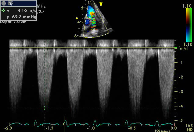
- Pulmonary Stenosis
- Continuous wave spectral Doppler across the pulmonary valve in a patient with severe pulmonary valve stenosis
- Echocardiographic Assessment: Parasternal Short Axis (Pulmonary Valve)
- Assess the pulmonary valve for dysplasia (leaflet morphology, excursion, hypoplasia, tethering)
- Assess main pulmonary artery (hypoplasia, post-stenotic dilation in case of valvar PS)
- Assess branch pulmonary arteries (size, stenosis)
- Assess for pulmonary stenosis or regurgitation (color and spectral Doppler pulse wave and continuous wave Doppler) at the subvalvar, valvar, supravalvar level and interrogate branch pulmonary arteries
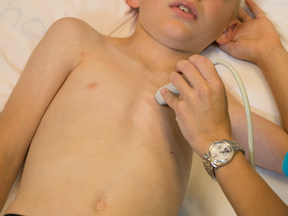
- Left sternal border
- 3rd or 4th intercostal space
- Notch pointing towards the left shoulder (1-2 o'clock)
- Transducer tilted superiorly and medially