- Normal appearing mitral valve leaflets with good excursion
- Normal caliber left ventricular outflow tract without narrowing
- Normal appearing aortic valve leaflets with good excursion
- Normal left ventricular systolic function
- Normal appearing aortic root without dilation
Normal lamianr flow across the left ventricular outflow tract and aortic valve
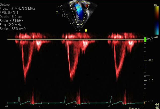
- Normal pulse wave spectral Doppler across the aortic valve
- No aortic stenosis (defined as a peak velocity <2.0 meters/sec)
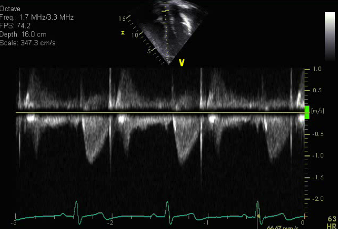
- Normal continuous wave spectral Doppler across the aortic valve
- No evidence of left ventricular outflow tract obstruction (defined as a peak velocity <2.0 meters/sec)
- No aortic valve stenosis (defined as a peak velocity <2.0 meters/sec)
- No supravalvar aortic stenosis (defined as a peak velocity <2.0 meters/sec)
The apical five chamber view is an excellent view to assess the left ventricular outflow tract and flow across the aortic valve.
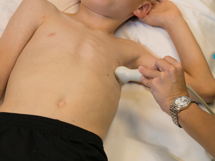
- Transducer at cardiac apex
- Notch at 3-4 o'clock (slight clockwise rotation)
- 4th or 5th intercostal space
- Probe tilted anteriorly (tail down)
- Aortic stenosis
- Subaortic membrane
- Hypertrophic cardiomyopathy with septal hypertrophy
- Aortic valve stenosis
- Supravalvar aortic stenosis
- Mitral valve abnormality
- Mitral valve prolapse
- Supravalvar mitral ring
- Mitral valve arcade
- Parachute mitral valve