- Aortic valve appears to have thin leaflets with normal systolic excursion
- Normal appearing aortic root, sinotubular junction and ascending aorta
- Normal laminar flow across subaortic, aortic valve and supravalvar aortic region
- No aortic regurgitation
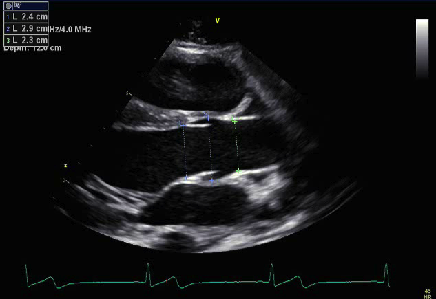
Still frame measurement of aortic dimensions
- Aortic valve
- Aortic root
- Sinotubular junction
This parasternal long axis view focuses on the left ventricular outflow tract, aortic valve, aortic root, sinotubular junction and ascending aorta.
.png)
- Transducer placed at the left sternal border
- 3rd or 4th intercostal space
- Notch pointed towards the patient's right shoulder (11 o'clock)
- Ultrasound beam zoomed or sector width narrowed to focus on aortic valve, root, STJ and ascending aorta
- Aortic root dilation or aneurysm (Marfan Syndrome)
- Ascending aortic dilation (bicuspid aortic valve)
- Aortic regurgitation or stenosis
- Subaortic membrane