- Normal caliber aortic arch and head and neck vessels
- No evidence of coarctation of the aorta (narrowing in region of aortic isthmus)
Normal laminar flow in the aortic arch and head and neck vessels
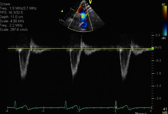
- Normal pulse wave Doppler in the descending aorta
- No evidence of arch obstruction in the region sampled in the descending aorta (defined as a peak velocity <2.0 meters/sec)
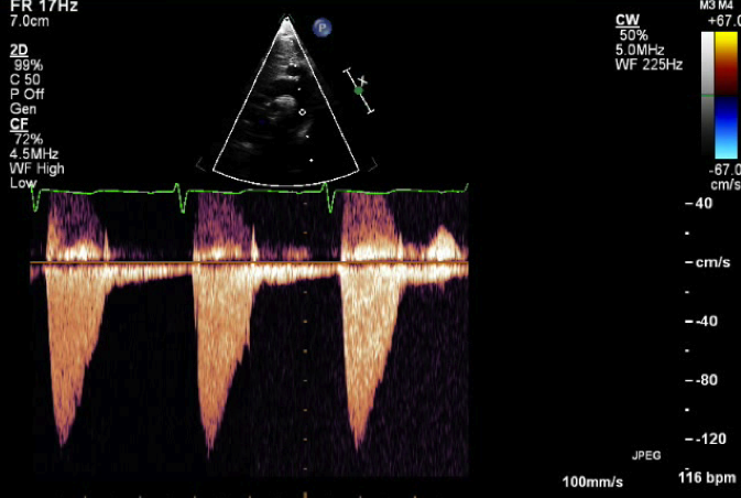
- Normal continuous wave Doppler pattern in the descending aorta
- No evidence of obstruction in the descending aorta (defined as a peak velocity<2.0 meters/sec)
- Color compare image of aortic arch
- Normal caliber aortic arch without evidence of narrowing or coarctation of the aorta
- Normal laminar flow in the ascending, transverse and descending thoracic aorta
This view obtained from the suprasternal notch is best for profiling the ascending aorta, transverse arch, aortic isthmus and descending aorta. In addition, the head and neck vessels (carotid and subclavian arteries) can be visualized from this view.
.png)
- Probe placed in suprasternal notch
- Notch pointed towards 1 o'clock
- Tilt inferiorly and anteriorly
- Coarctation of the aorta
- Aortic arch hypoplasia
- Ascending aortic dilation
- Good view for spectral Doppler to quantify aortic stenosis (valvar and supravalvar)
- Head and neck vessel abnormalities or variants
- Common brachiocephalic trunk