- Normal sized right coronary artery ostia and proximal right coronary artery
- No evidence of coronary artery dilation or aneurysm
- Color compare view of 2D and color in the proximal right coronary artery
- The coronary artery fills normally in diastole (red flow at coronary ostium in diastole)
- Normal appearing right coronary artery ostia and proximal right coronary artery
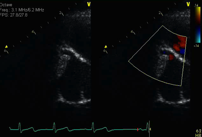
- Sill image of right coronary artery
- Normal flow into the proximal right coronary artery is seen during diastole
This view obtained from a parasternal short axis view is one of the ideal views for profiling the origin, caliber and course of the right coronary artery.
-
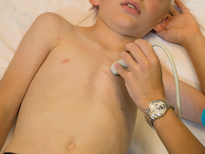
Transducer placed at left sternal border
-
3rd or 4th intercostal space
-
Notch pointed towards patient's left shoulder (1-2 o'clock)
-
Probe in neutral position
-
Slight counterclockwise rotation if needed
-
Anomalous right coronary artery
-
Right coronary artery dilation or aneurysm