- Normal caliber main and branch pulmonary arteries
- The branch pulmonary arteries appear confluent and symmetric in size
- Color compare view the the main and branch pulmonary arteries
- Normal size main pulmonary artery
- Normal size and confluent branch pulmonary arteries
- Normal laminar flow in the main and branch pulmonary arteries
- No evidence of aortopulmonary shunting (patent ductus arteriosus or aortopulmonary window)
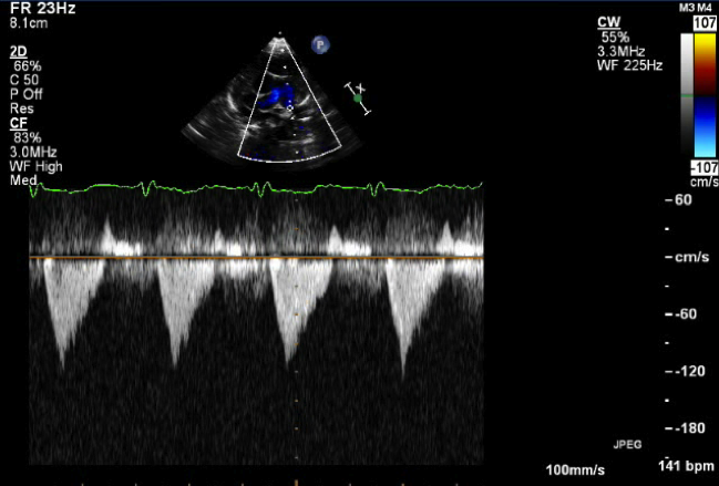
Continuous wave Doppler in the left pulmonary artery reveals no evidence of obstruction (defined as a peak velocity <2.0 meters/sec)
Also known as the "pants view", this view obtained from the high parasternal short axis can profile the distal main pulmonary artery and branch pulmonary arteries.

- Modified parasternal short axis view (slightly modified view from picture above)
- Left sternal border
- 3rd or 4th intercostal space (slide upward 1-2 rib spaces and slightly more medial to obtain this modified view)
- Notch pointing towards 3 o'clock
- Branch pulmonary artery dilation, hypoplasia or stenosis
- Main pulmonary artery dilation, hypoplasia or stenosis
- Aortopulmonary window
- Patent ductus arteriosus (PDA)