- Normal caliber pulmonary veins
- Two right and two left pulmonary veins appear to connect normally to the left atrium
- Normal laminar flow demonstrated in the pulmonary veins as they drain into the left atrium
- Lower pulmonary veins (red flow)
- Upper pulmonary veins (blue flow)
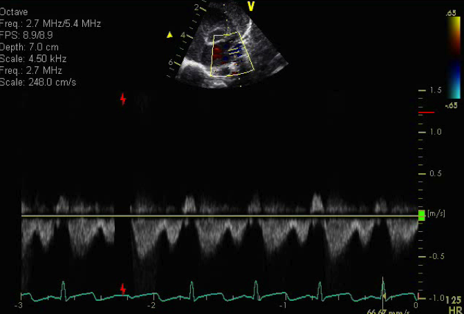
- Normal triphasic pulse wave spectral Doppler pattern sampled from the left upper pulmonary vein
- Pulmonary venous systolic wave (S)
- Pulmonary venous early diastolic wave (D)
- Pulmonary venous atrial reversal flow wave (A)
This view obtained from the suprasternal notch is best for profiling all of the individual pulmonary veins as they drain into the left atrium. Each vein should always be assessed by both color and spectral Doppler to confirm normal connection and unobstructed drainage.
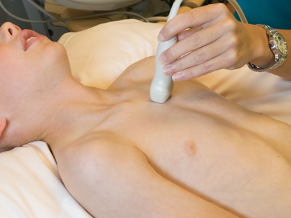
- Probe at suprasternal notch
- Notch at 3 o'clock
- Transducer tilted posteriorly (tail up)
- May have to slide probe inferiorly along the left side of sternum with a posterior inferior tilt of the transducer (tail up) to fully profile pulmonary veins
- Left superior vena cava
- Pulmonary vein stenosis
- Anomalous pulmonary veins
- Total anomalous pulmonary venous return
- Partial anomalous pulmonary venous return