- Normal caliber IVC
- The IVC appears to connect normally uninterrupted to the right atrium
- Normal laminar flow from the hepatic veins and IVC into the right atrium
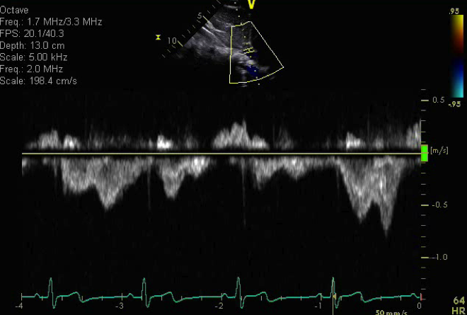
Normal pulse wave Doppler in the IVC
This view of the IVC obtained from the subcostal view is an ideal view to profile the inferior vena cava and hepatic veins.
.png)
- Probe position in subxyphoid location
- Rotate probe so notch at 12 o'clock
- Tilt to patient's left
- Inferior vena cava (IVC) abnormality
- Interrupted IVC
- IVC narrowing
- IVC thrombosis or vegetation
- Hepatic venous drainage abnormality