- Normal systemic venous return to the right atrium (superior vena cava and inferior vena cava)
- Intact atrial septum without evidence of atrial septal defects
- Normal laminar flow from the superior (red flow) and inferior (blue flow) vena cava into the right atrium
- The atrium septum appears intact without evidence of shunting across the atrial septum
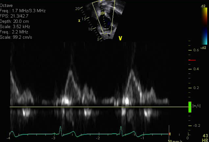
Normal pulse wave Doppler in the superior vena cava
This subcostal short axis (also known as subcostal saggital view) is an excellent view to profile the SVC and IVC and atrial septum.
.png)
- Transducer on abdomen just below xyphoid process
- Notch at 6 o'clock
- Probe tilted towards patient's right shoulder
- Atrial septal defects
- Superior vena cava narrowing or stenosis
- Anomalous pulmonary venous drainage
- Right atrial enlargement
- Interrupted inferior vena cava